Article from Autism Speaks March 26, 2014
Original Study from New England Journal of Medicine Patches of Disorganization in the Neocortex of Children with Autism research
Researchers find common pattern of disruption to prenatal brain development; propose that early intervention may help brain compensate
Researchers say they’ve found clear and direct evidence that autism begins during prenatal brain development.
A new study provides physical evidence that autism spectrum disorder (ASD) has roots in prenatal development. The research, supported in part by Autism Speaks, appears online in the New England Journal of Medicine.
The researchers studied the donated post-mortem brain tissue of 11 children who had been diagnosed with autism and of 11 unaffected children. In 10 out of the 11 ASD cases, they found recurring patches of abnormal development in layers of the cerebral cortex that form during prenatal development. By contrast, they found the patches in just 1 of the 11 children unaffected by autism.
“The finding that these defects occur in patches rather than across the entirety of cortex gives hope as well as insight,” says senior author Eric Courchesne, of the University of California, San Diego. Early intervention therapies for autism may help children’s brains “rewire” to circumvent the defective patches, Dr. Courchesne proposes. Further research may advance understanding of how to best foster such improvement.
“This study is a prime example of how precious tissue donations, technological advances in neuroscience tools and a lot of hard work by talented researchers can shine new light onto the biological roots of autism,” comments Dan Smith, Autism Speaks senior director of discovery neuroscience. “Numerous brain imaging studies have revealed that ASD can affect how the brain functions. But this study takes us to a new level by homing in on early life changes in the brain’s fundamental building blocks.” Dr. Smith was not directly involved in the research.
The physical evidence of early brain changes also backs a growing body of research linking increased risk for autism to genetically controlled processes and environmental influences that affect prenatal brain development.
A unique pattern of prenatal changes ”The most surprising finding was the similar early developmental pathology across nearly all of the autistic brains,” says co-author Ed Lein, “especially given the diversity of symptoms in those with autism, as well as the extremely complex genetics behind the disorder.”
The team found abnormal patches across the brain’s frontal cortex and temporal cortex. The frontal cortex is associated with complex communication and social abilities. The temporal cortex is associated with language.
By contrast, the researchers found no abnormalities in the visual cortex. They note that visual perception tends to be unimpaired – sometimes even heightened – in individuals with autism.
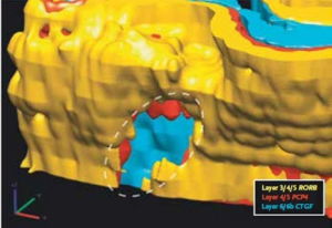
The study also produced a three-dimensional brain model of the patches that failed to develop the normal cell-layering pattern.

Three-D reconstruction of a patch defect (dashed circle) found scattered through certain prenatally formed layers of brain tissue from children affected by autism.
During early brain development, each cortical layer develops a specific brain-cell type with specific patterns of connectivity that perform unique roles in processing information, Dr. Courchesne explains. In the process, each brain-cell type and tissue layer acquires a distinct genetic signature. The researchers used these genetic markers – and their absence – to build their 3-D model of normally developing and abnormally developing brain layers.
The donated brain tissue making this study possible came, in part, from the Autism Speaks Autism Tissue Program. The program is currently becoming part of a new collaborative tissue bank called BrainNet.
“It’s so important to emphasize the critical role of donated brain tissue in enabling researchers to find answers to questions about brain development,” says Autism Speaks Chief Science Officer Rob Ring. “Tissue-based research like this can happen only when families make the ultimate donation in the face of tragedy. Through their generosity, we can help ensure that these resources remain available for breakthrough research like this.”
Autism Speaks is currently funding follow-up research on the team’s discoveries through a Meixner Postdoctoral Fellowship in Translational Research for Haim Bellinson, at the University of California, San Francisco. The study published today was supported by a research grant for Dr. Courchesne from Autism Speaks, as well as funding from the Simons Foundation, the Peter Emch Family Foundation, the Thursday Club Juniors, the UCSD Autism Center of Excellence and the Allen Institute for Brain Science.
Enter the text or HTML code here

